
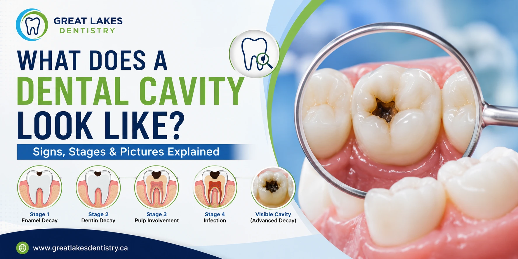
Dental cavities progress through distinct stages — catching them early is the key to simpler, less costly treatment
What Does a Dental Cavity Look Like? Signs, Stages & Pictures Explained
Most people assume they would know if they had a cavity — but the truth is that dental cavities are often completely invisible and painless in their early stages. By the time a cavity becomes obvious to the naked eye or starts causing discomfort, it has usually progressed far enough to require more invasive treatment. Understanding what a dental cavity looks like at each stage of development — and what signs to watch for before it becomes visible — is one of the most practical things you can do for your long-term oral health.
What Is a Dental Cavity?
A dental cavity — clinically called dental caries — is a permanently damaged area in the hard surface of a tooth that develops into a hole or pit. Cavities form when bacteria in the mouth produce acids from the sugars and carbohydrates in food and drink. Those acids gradually dissolve the mineral structure of the tooth, progressing from the outer enamel layer inward toward the dentin and, if untreated, the pulp (nerve) at the tooth’s core.
Cavities are among the most common chronic health conditions in the world. According to the World Health Organization, dental caries affects nearly 2.5 billion people globally — making understanding and prevention more important than ever.
Crucially, cavities do not heal on their own once they have progressed past the initial demineralization stage. The damage is permanent and requires professional treatment to stop.
What Does a Dental Cavity Look Like? Stage by Stage
Stage 1: Demineralization — White Spot Lesions
What it looks like: A chalky white or off-white spot on the tooth surface, most commonly near the gum line or between teeth.
This is the earliest visible sign of cavity formation — and the only stage where the process may be reversible without a filling. Demineralization occurs when acid erodes the mineral content of the enamel surface without yet breaking through it. The affected area loses its translucent sheen and appears white or cloudy compared to surrounding healthy enamel.
At this stage, fluoride treatments, improved oral hygiene, and dietary changes can potentially remineralize the lesion and halt cavity progression. This is why regular professional teeth cleaning and checkup appointments — where early white spot lesions can be identified and treated — are so critical.
Patient experience: Usually no pain or sensitivity at this stage. Most patients are completely unaware a white spot lesion exists without a dental examination.
Stage 2: Enamel Decay — Surface Cavity
What it looks like: A brown, black, or dark spot on the tooth surface. On smooth surfaces, it may appear as a discoloured patch. On chewing surfaces, it may look like a small dark pit or groove.
Once acid erosion breaks through the enamel surface, a true cavity has formed. The outer enamel layer has been irreversibly damaged. The discolouration occurs as bacteria and organic debris accumulate in the cavity opening.
What it looks like by location:
- Chewing surfaces (occlusal) — dark brown or black spots in the pits and fissures of molars and premolars. These are among the most common cavity locations because food particles collect easily in these grooves
- Between teeth (interproximal) — not visible to the naked eye; only detectable with X-rays. This is why professional X-rays are essential even when no visible decay is present
- At the gum line (cervical) — yellowish-brown discolouration where the tooth meets the gum. Common in patients with gum recession or dry mouth
- On smooth surfaces — brown or dark patches on the cheek-side or tongue-side of teeth
Patient experience: Mild to moderate sensitivity to sweet foods and cold drinks may begin at this stage. Some patients experience brief sharp pain when eating sugary foods.
Stage 3: Dentin Decay — Cavity Progresses Inward
What it looks like: A larger, darker cavity — often visibly brown or black. The cavity may appear hollow or pit-like. Surrounding enamel may appear grey or undermined.
Once decay reaches the dentin — the softer, yellowish layer beneath the enamel — it progresses significantly faster. Dentin contains tiny fluid-filled tubules that connect to the tooth’s nerve, which is why dentin-level cavities cause noticeably more sensitivity and pain than surface cavities.
Patient experience: Increased sensitivity to hot, cold, and sweet. Lingering ache after eating. The cavity may be large enough to be felt with the tongue. At this stage, treatment is a filling — typically a dental crown may be recommended if the cavity is extensive enough to compromise the tooth’s structural integrity.
Stage 4: Pulp Involvement — Abscess Risk
What it looks like: A large, visibly dark cavity — often covering a significant portion of the visible tooth surface. The tooth may appear grey, darkened, or structurally compromised. In some cases, a visible hole or broken edge is present.
When decay reaches the pulp — the innermost chamber containing the tooth’s nerves and blood vessels — the infection triggers an inflammatory response. This is the stage at which patients typically experience severe, spontaneous pain, and where the risk of a dental abscess becomes significant.
Patient experience: Severe, throbbing, spontaneous pain — often worse at night when lying down. Pain that lingers long after eating or drinking. Possible visible swelling of the gum or face. A dark, visibly broken or crumbling tooth. Treatment at this stage typically requires root canal treatment or extraction. If you are experiencing these symptoms, our emergency dentist team at Great Lakes Dentistry is available for same-day assessment.
Stage 5: Tooth Loss — Advanced Decay
What it looks like: A severely broken-down tooth, often reduced to a dark stump or root fragment at the gum line. Large portions of the visible tooth may be missing entirely.
At the most advanced stage of untreated decay, the tooth structure has been so extensively damaged that restoration may no longer be possible. Extraction is typically the only remaining option — followed by tooth replacement planning.
Patient experience: Often less acute pain at this stage, as the nerve may have died — but chronic infection, abscess, and bone loss are common. Many patients at this stage experience recurrent swelling, bad taste, and foul odour from the affected tooth.
Where Do Cavities Most Commonly Form?
Understanding which areas of the mouth are most vulnerable helps you prioritize your cleaning routine:
Highest Risk Areas
The pits and fissures of back molars and premolars (chewing surfaces). Between teeth (interproximal) — only detectable by X-ray. Along the gum line, particularly in patients with gum recession. Around the edges of existing fillings, crowns, or other restorations.
Why These Areas Are Vulnerable
Deep grooves trap food and bacteria that brushing cannot always reach. Between-teeth surfaces are missed entirely by a toothbrush — only floss reaches them. Gum recession exposes the softer root surface (cementum) which decays faster than enamel. Old restorations develop micro-gaps over time where bacteria re-enter.
Signs You May Have a Cavity — Even Without a Visible Hole
Because many cavities are not visible to the naked eye, watching for these clinical signs is important:
- Tooth sensitivity — a sharp, brief pain when consuming cold drinks, sweet foods, or hot beverages
- Spontaneous toothache — pain that occurs without an obvious trigger, particularly at night
- Pain when biting — discomfort specifically when chewing or biting down on the affected tooth
- Visible discolouration — brown, black, or white spots on any tooth surface
- Bad breath that doesn’t resolve — bacterial activity in a cavity produces odour-causing compounds
- A rough, sticky, or sharp-edged feeling when running your tongue over a tooth surface
- Food consistently getting stuck in the same spot between teeth
How Are Cavities Diagnosed?
At Great Lakes Dentistry, cavity detection involves several tools used together for maximum accuracy:
- Visual examination — using magnification and dental lighting to identify visible decay, discolouration, and structural changes
- Digital X-rays — essential for detecting interproximal (between-teeth) cavities and sub-surface decay not visible to the eye. Digital X-rays expose patients to significantly less radiation than traditional film X-rays
- Tactile exploration — a dental explorer instrument detects soft, sticky spots in enamel that indicate demineralization or early decay
- Laser cavity detection — in some cases, laser fluorescence devices help identify decay in fissures before it becomes radiographically visible
What Happens If a Cavity Is Left Untreated?
Untreated cavities do not stabilize — they always progress. The consequences of delayed treatment escalate significantly at each stage:
- A Stage 1 white spot lesion treated with fluoride costs nothing beyond a checkup
- A Stage 2 enamel cavity requires a simple filling
- A Stage 3 dentin cavity may require a larger filling or crown
- A Stage 4 pulp-involved cavity requires root canal treatment — then a crown
- A Stage 5 advanced cavity typically requires extraction and then tooth replacement with a dental implant or bridge
The cost — financially and in terms of treatment complexity — increases at every stage. Early detection is always the most affordable, least invasive option.
How to Prevent Dental Cavities
The good news is that cavities are almost entirely preventable with the right habits:
- Brush twice daily for two full minutes with a fluoride toothpaste — paying close attention to the gum line and back molars
- Floss once daily — the only way to clean between teeth where the majority of interproximal cavities form
- Reduce sugar and acid exposure — limit sugary snacks and drinks, and rinse with water after consuming them
- Drink fluoridated water — municipal water fluoridation remains one of the most effective public health interventions for cavity prevention
- Ask about dental sealants — thin protective coatings applied to the chewing surfaces of back molars are highly effective at preventing pit-and-fissure cavities, especially in children and teens
- Schedule regular checkups — the Canadian Dental Association recommends checkups every six months for most patients
According to the Canadian Dental Association, consistent at-home oral hygiene combined with regular professional care is the most effective strategy for preventing dental caries at every age.
Trusted Resources on Dental Cavities
- Canadian Dental Association — Oral Health Resources
- Health Canada — Dental Care Guidelines
- World Health Organization — Oral Health Facts
Think You Might Have a Cavity? Book a Checkup at Great Lakes Dentistry, Oakville
The earlier a cavity is caught, the simpler and more affordable the treatment. Call Dr. Gurpreet Mohi and our team at (905) 825-1212 or book online for a comprehensive exam with digital X-rays.
Frequently Asked Questions: What Does a Dental Cavity Look Like?
What does a cavity look like on a tooth?
Cavities appear differently depending on their stage and location. Early-stage cavities appear as chalky white spots on the enamel surface. As they progress, they develop into brown, dark brown, or black spots or pits — particularly visible on the chewing surfaces of back teeth. Advanced cavities may look like large dark holes, crumbling tooth structure, or a visibly broken-down tooth. Many cavities, particularly between teeth, are completely invisible without dental X-rays.
Can I see a cavity on my tooth at home?
You may be able to see a cavity on visible tooth surfaces if it has progressed to the enamel or dentin stage — particularly on the chewing surfaces or front of back teeth. However, many of the most common cavities form between teeth where they are completely invisible during self-examination. Only a dentist using X-rays and clinical instruments can reliably detect all cavities, including early-stage lesions that appear completely normal to the naked eye.
Do all cavities hurt?
No — and this is one of the most important misconceptions about cavities. Early-stage cavities (demineralization and enamel decay) cause no pain at all. Pain typically only begins when decay reaches the dentin layer, and becomes severe when the pulp (nerve) is involved. Waiting for pain before seeking dental care means waiting until the cavity has progressed to a more advanced, more costly, and more invasive stage of treatment.
What is the black spot on my tooth — is it always a cavity?
Not necessarily. A black or dark spot on a tooth can indicate active decay, but can also be surface staining from coffee, tea, wine, or tobacco — or a stained groove that is not yet decayed. The difference is clinically significant and requires professional diagnosis. A dentist can distinguish between a stain and active decay using visual examination, dental instruments, and X-rays. Never assume a dark spot is harmless without having it assessed.
Can a cavity heal on its own?
In the very earliest stage — demineralization, or white spot lesions — the enamel can potentially remineralize with fluoride treatment, improved oral hygiene, and dietary changes. However, once a true cavity has formed (once the enamel surface has been broken through), the damage is permanent and cannot reverse without professional treatment. There is no home remedy that will fill or repair an established cavity.
How quickly does a cavity progress?
The rate of cavity progression varies depending on diet, oral hygiene habits, saliva flow, fluoride exposure, and individual bacterial composition. A cavity in enamel may remain stable for months or years with good hygiene. Once decay reaches dentin, progression accelerates significantly — sometimes progressing to pulp involvement within months if untreated. This variability is why regular six-month checkups — rather than waiting for symptoms — are the standard of care.
What does a cavity between teeth look like?
Interproximal cavities (between teeth) are invisible to the naked eye in their early stages — they form on the contact surfaces of adjacent teeth that touch each other. On dental X-rays, they appear as a dark triangular or wedge-shaped shadow near the contact point. Some patients only notice an interproximal cavity when it becomes large enough to cause food trapping, sensitivity, or visible darkening near the gum line between teeth.
How often should I get checked for cavities?
The Canadian Dental Association recommends dental checkups every six months for most healthy adults and children. Some patients with a history of frequent cavities, dry mouth, high sugar intake, or gum disease may benefit from more frequent visits — every 3 to 4 months. Your dentist will advise on the frequency that is most appropriate for your individual risk profile. Consistency is key — gaps in care allow cavities to progress undetected.
Final Thoughts
Key Takeaways: What Does a Dental Cavity Look Like?
- Cavities progress through five distinct stages — from invisible white spot lesions to visible dark holes and eventual tooth loss
- Early-stage cavities cause no pain and are invisible without professional examination and X-rays
- White spots on enamel are the first visible sign — and the only stage where progression may be reversible
- Brown, black, or dark spots on tooth surfaces typically indicate active or progressed decay requiring treatment
- Between-teeth cavities are only detectable with X-rays — another reason regular checkups are essential
- The earlier a cavity is treated, the simpler and more affordable the procedure — always